


What Is Ultrasound Imaging?
Οι υπέρηχοι (υπερηχογραφήματα) υπάγονται στην κατηγορία των πιο αξιόπιστων και ασφαλών απεικονιστικών μεθόδων στη σύγχρονη ιατρική. Είναι μια κατηγορία διαγνωστικών εξετάσεων που μπορεί να προταθεί σε πολλές περιπτώσεις ώστε να γίνει διάγνωση που αφορά διάφορα σημεία και όργανα του σώματος. Για την διεξαγωγή των υπερηχογραφημάτων, χρησιμοποιούνται ηχητικά κύματα υψηλών συχνοτήτων για να απεικονιστεί το μέγεθος, το σχήμα και η δομή των εσωτερικών οργάνων και των αιμοφόρων αγγείων υπό την καθοδήγηση εξιδεικευμένου ιατρού.
Πρόκειται για μια ανώδυνη και ακίνδυνη μέθοδο η οποία διεξάγεται σε σύντομο χρόνο και έχει χαμηλό κόστος. Με την βοήθεια της τεχνολογίας σήμερα μπορούμε να πετύχουμε διάγνωση ακριβείας σε πλήθος παθολογικών καταστάσεων.
Ο εξοπλισμός The Ultrasound Imaging Department is equipped with state-of-the-art, highly reliable, and advanced technology systems.
Ultrasound Imaging
Our Ultrasound Department offers a wide range of diagnostic examinations for both adults and children (from the age of 3 years onwards).
The Ultrasound Imaging Department is equipped with state-of-the-art, highly reliable, and advanced medical technology.
• Upper Limb Arterial and Venous Duplex Ultrasound
The arteries and veins of the upper limbs are examined to assess vessel patency and identify possible stenosis or thrombosis. Pre-operative vascular mapping is performed for the creation of an arteriovenous fistula (AV fistula). In patients undergoing haemodialysis, the fistula or vascular graft is evaluated to confirm patency, detect possible complications, and calculate blood flow volume, which is essential for effective haemodialysis treatment.
Lower Limb Arterial and Venous Duplex Ultrasound
The arteries and veins of the lower limbs are examined to assess vessel patency and identify possible stenosis or thrombosis. The examination also evaluates the presence of superficial venous insufficiency, varicose veins, and subcutaneous oedema of the lower limbs.
Carotid and Vertebral Artery Duplex Ultrasound
The arteries supplying blood to the brain (carotid and vertebral arteries) are examined to assess vessel patency, intima-media thickness, and the possible presence of atherosclerotic plaques. The degree of internal carotid artery stenosis is calculated, while the echogenic characteristics of atherosclerotic lesions are evaluated and classified as calcified, soft, or mixed plaques. The examination also assesses for possible subclavian steal syndrome and arterial dissection.
Abdominal Aorta and Iliac Artery Duplex Ultrasound
The abdominal aorta and/or iliac arteries are examined to assess vessel patency, atherosclerotic changes, thrombosis, and luminal diameter. The examination also evaluates for the possible presence of aneurysmal dilatation, aneurysm formation, or arterial dissection.
Renal Artery Duplex Ultrasound
The main renal arteries are visualised along their course towards the kidneys, together with their intrarenal branches, in order to assess renal blood supply and exclude renal artery stenosis. In kidney transplant recipients, the examination evaluates the patency of the renal artery and the adequacy of vascular perfusion to the transplanted graft.
Upper and Lower Abdominal Ultrasound (U/S)
The solid abdominal organs examined include the liver, gallbladder, biliary ducts, pancreas, spleen, kidneys, urinary bladder, uterus, ovaries, and prostate gland. These organs are assessed in terms of their size, texture, and the possible presence of pathology, such as focal lesions, gallstones or urinary calculi, dilatation of the bile ducts or renal collecting system, prostatic enlargement, urinary retention, and other abnormalities. Organs containing air, such as the stomach, small intestine, and large intestine, are generally not well visualised on ultrasound, as air interferes with the transmission of ultrasound waves. However, in specific cases (such as Crohn’s disease), targeted examination of bowel loops may be performed.
Thyroid Ultrasound (U/S)
The thyroid gland is examined to assess its size and echotexture, as well as the possible presence of thyroid nodules. The examination is also used for the monitoring of chronic thyroid conditions, such as Hashimoto’s disease. In patients who have undergone thyroidectomy, the scan evaluates for any residual thyroid tissue or abnormal lymph nodes. The parathyroid glands are also assessed when there is suspicion of a parathyroid adenoma.
Transvaginal Ultrasound (U/S)
The uterus and ovaries are examined in greater detail compared with a standard transabdominal (suprapubic) pelvic ultrasound. The examination assesses their size and echotexture, as well as the possible presence of cystic or solid lesions.
Soft Tissue Ultrasound (U/S)
The examination is tailored according to the patient’s symptoms and the anatomical area of concern. It may include assessment for conditions such as inguinal hernia, umbilical hernia, palpable subcutaneous masses, axillary lymph nodes, and other soft tissue abnormalities.
Scrotal Ultrasound (U/S)
The testes and epididymides are examined to assess their size, echotexture, and the possible presence of focal lesions. The examination also evaluates testicular vascularity and the possible presence of varicocele. In addition, abnormal fluid collection within the scrotum (hydrocele) or the presence of bowel loops extending into the scrotum (inguinoscrotal hernia) can also be identified.
Specialised Ultrasound Examinations & Image-Guided Needle Procedures
Liver Elastography
Liver elastography is a modern, non-invasive diagnostic examination used to assess the degree of hepatic fibrosis and detect the possible presence of liver cirrhosis. The examination is performed using a latest-generation ultrasound system equipped with shear wave elastography technology. This advanced technique enables simultaneous visualisation of the liver and measurement of its elasticity by recording the propagation speed of shear waves generated within the tissues following acoustic pulse-induced deformation. Based on these measurements, it is possible to determine the stage of liver fibrosis and identify the presence of cirrhosis.
Thyroid Elastography
Thyroid elastography is an imaging technique used to quickly and painlessly assess the stiffness and elasticity of thyroid nodules and thyroid parenchyma. It is performed as a complementary examination to thyroid ultrasound and provides additional diagnostic information for the evaluation of thyroid disorders.
• Ελαστογραφία μαστού
Η Ελαστογραφία μαστού, είναι μια νέα υπερηχογραφική τεχνική που χρησιμοποιείται προκειμένου να εκτιμήσει το βαθμό σκληρότητας μιας βλάβης του μαστού. Εξετάζει τις μηχανικές και ελαστικές ιδιότητες των μαλακών ιστών. Ορισμένες παθολογικές καταστάσεις προκαλούν σημαντικές αλλαγές στη δομή των μαλακών μορίων, τροποποιώντας τις ελαστικές τους ιδιότητες και οδηγώντας σε αυξημένη σκληρότητα.
Η Ελαστογραφία είναι μια νέα υπερηχογραφική τεχνική απεικόνισης που μπορεί να βοηθήσει στη διαφορική διάγνωση μιας καλοήθους από κακοήθη μάζα στον μαστό με βάση τον βαθμό σκληρότητάς της. Η ελαστογραφία χρησιμοποιείται στον μαστό:
• Προκειμένου να διερευνηθεί κάποια μικρή βλάβη που ανιχνεύεται με το υπερηχογράφημα αλλά δεν απεικονίζεται στην μαστογραφία, έτσι ώστε να ακολουθήσει βιοψία αντί της παρακολούθησης με στόχο τον εντοπισμό πρώιμων μικρών κακοηθειών.
• Σε βλάβες που έχουν χαρακτηριστεί από το κλασσικό υπερηχογράφημα σαν μάλλον καλοήθεις, ώστε σε περίπτωση που και η ελαστογραφία έχει καλοήθη χαρακτηριστικά να υπάρξει αραίωση στα διαστήματα παρακολούθησης
• Σε βλάβες που έχουν χαρακτηριστεί από το κλασσικό υπερηχογράφημα σαν μάλλον κακοήθεις, ακόμα και αν τα χαρακτηριστικά της βλάβης με την ελαστογραφία είναι καλοήθη τότε οι βλάβες αυτές πρέπει να διερευνώνται με βιοψία.
UGAP (Ultrasound-Guided Attenuation Parameter – Assessment of Hepatic Steatosis)
UGAP is used for the evaluation of Non-Alcoholic Fatty Liver Disease (NAFLD). It is particularly useful in overweight or obese individuals, as well as in patients with fatty liver infiltration, providing a quantitative assessment of liver fat content. The examination provides objective measurements expressed in dB/cm/MHz and enables grading of hepatic steatosis as S1, S2, or S3. It is performed for initial evaluation, establishment of a baseline reference for future monitoring, and prevention of progression to liver cirrhosis.
Ultrasound-Guided Fine Needle Aspiration (FNA) Biopsy of the Thyroid
Fine Needle Aspiration (FNA) biopsy of the thyroid gland is an effective diagnostic procedure performed to determine whether a thyroid nodule is benign or malignant. The procedure is relatively simple, minimally invasive, and painless, and usually takes less than 20 minutes to complete.
How Is Thyroid FNA Performed?
The procedure begins with a thyroid ultrasound examination, during which thyroid nodules are assessed for the likelihood of malignancy. Modern imaging techniques, including vascularity assessment and elastography, are utilised to evaluate the most suspicious features and identify the most appropriate area of the nodule for sampling. In addition, mapping of the surrounding cervical structures and lymph nodes is performed to investigate any possible signs of local infiltration or spread.
Breast Core Needle Biopsy (Core Biopsy)
Breast core needle biopsy is a procedure in which the physician obtains tissue samples from the breast using a specialised core biopsy needle. Representative tissue specimens are then sent for histopathological examination. If the findings are benign, the patient may avoid unnecessary surgical intervention. In cases where malignancy is identified, the physician can proceed with planning the appropriate surgical treatment and/or the corresponding medical therapy.
How Is a Breast Biopsy Performed?
The procedure begins with a breast ultrasound examination and evaluation of the imaging findings, followed by the administration of local anaesthesia. The core biopsy needle is then inserted through a very small incision in the skin of the breast. Tissue samples are subsequently obtained from the lesion or cystic area requiring biopsy and sent for histopathological analysis. The procedure takes approximately 15 minutes, is generally painless, and carries minimal risk of complications.
Cutting-Edge Technology
We utilise advanced echocardiographic imaging systems enhanced with Artificial Intelligence technology.
Our AI-powered algorithms reduce the need for manual processes and help optimise workflow efficiency through automated procedures, enabling faster, more accurate, and highly reliable diagnostic assessment.
With automated Cardiac Auto-Doppler and 2D LV measurements, we are able to reduce examination time while achieving maximum reproducibility and consistency of diagnostic results.
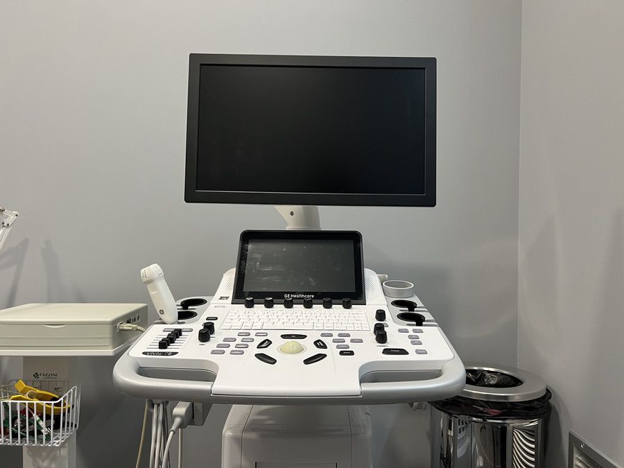
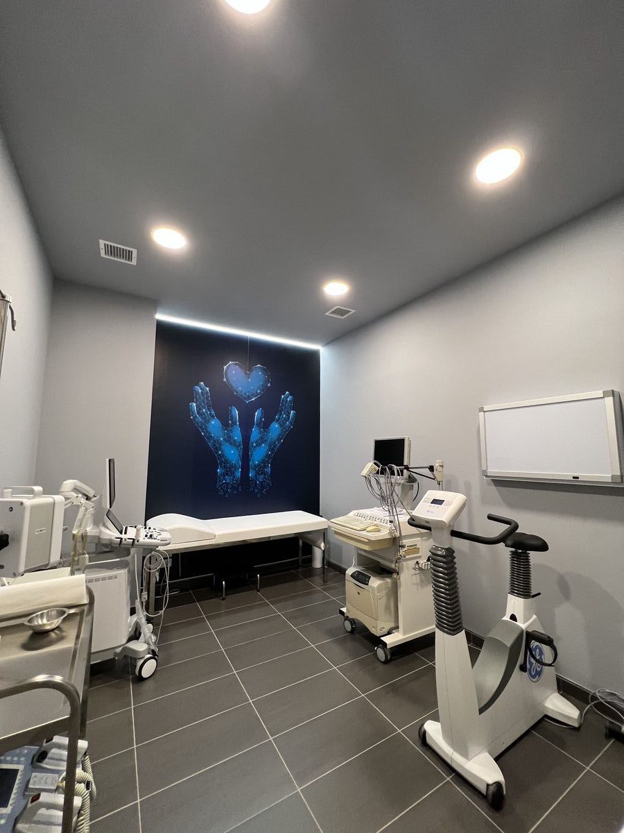
We use the GE Healthcare VIVID T8 Ultra Edition v206
A new-generation, versatile echocardiography platform with an innovative ergonomic design, combining high performance, exceptional imaging quality, and pioneering technologies and diagnostic techniques. The system supports both transthoracic and transoesophageal cardiac examinations, as well as advanced imaging applications for deep and peripheral vessels, upper and lower abdominal organs, small and superficial structures, obstetric and gynaecological imaging, paediatric and neonatal examinations, musculoskeletal imaging, urology, and transcranial applications.
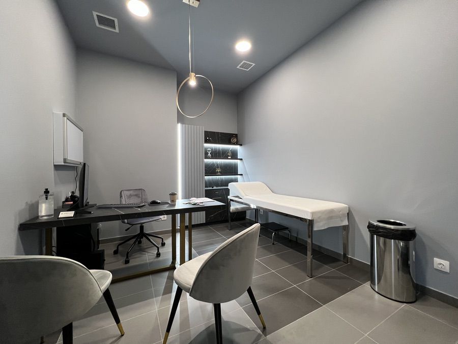
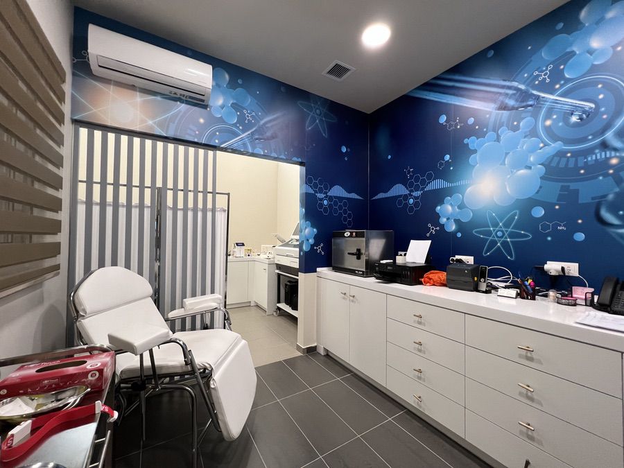
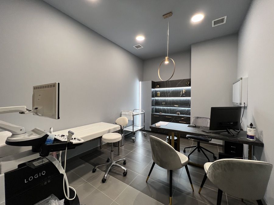
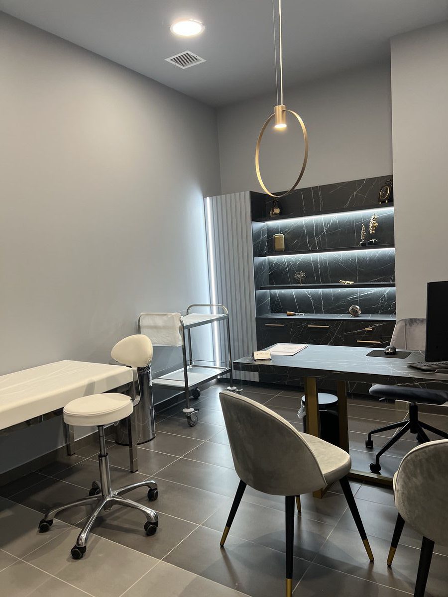
Get to Know Our Facilities
Frequently Asked Questions - Ultrasound Imaging
Results are typically available within approximately one hour, depending on the nature and complexity of the examination.
No harmful effects on humans have been identified from ultrasound examinations, as they do not use ionising radiation.
No, ultrasound examinations are completely painless.
We use the GE Healthcare VIVID T8 Ultra Edition v206
A new-generation, versatile echocardiography platform with an innovative ergonomic design, combining high performance, exceptional imaging quality, and pioneering technologies and diagnostic techniques. The system supports both transthoracic and transoesophageal cardiac examinations, as well as advanced imaging applications for deep and peripheral vessels, upper and lower abdominal organs, small and superficial structures, obstetric and gynaecological imaging, paediatric and neonatal examinations, musculoskeletal imaging, urology, and transcranial applications.
Interested?
Fill out the form and we’ll call you right away to provide more information.